Colloid Cysts In The Brain: Symptoms, Diagnosis, & Treatment
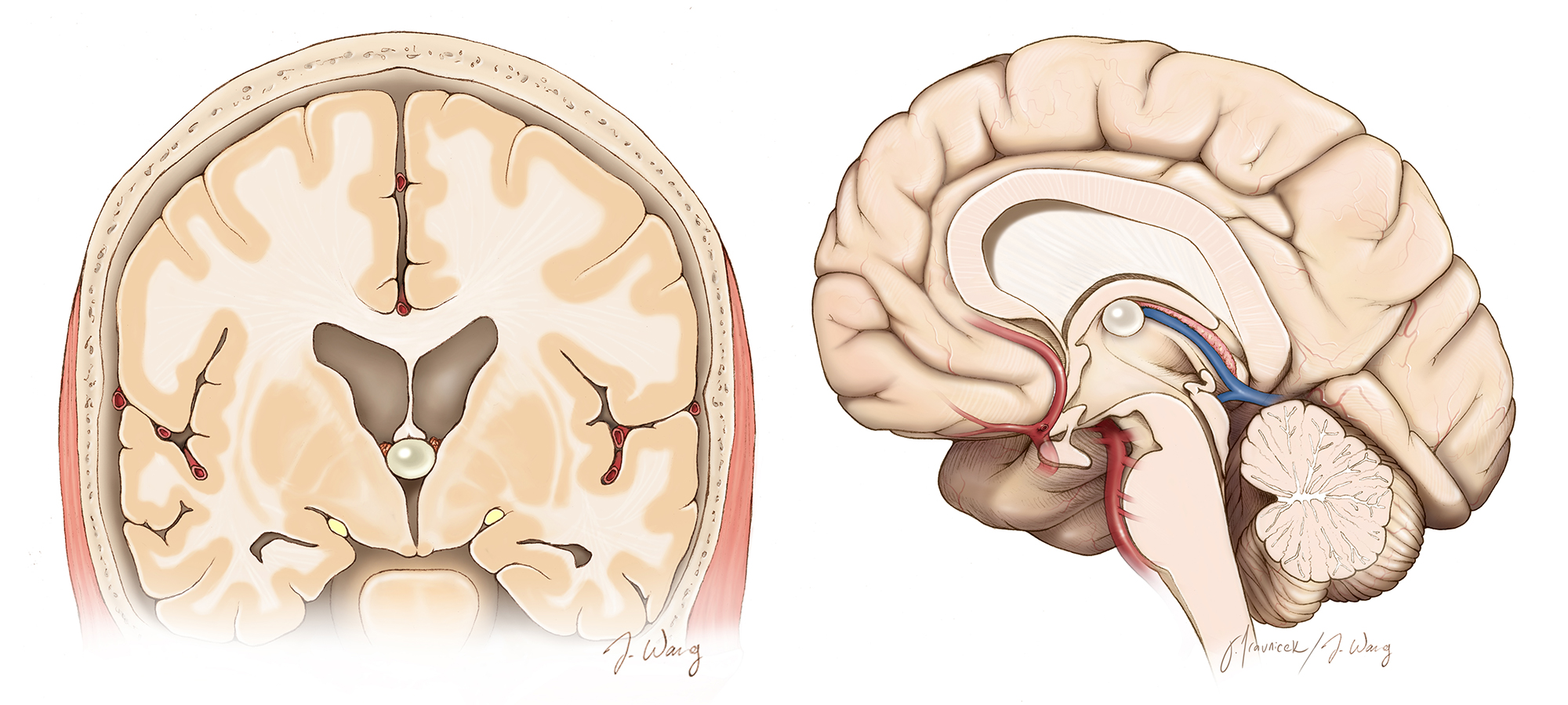

Colloid cysts, though rare, can have serious consequences due to their impact on CSF flow. Early diagnosis and prompt treatment are essential to prevent life-threatening complications. Colloid cysts are rare, benign lesions found in the third ventricle of the brain. Although these fluid-filled sacs are not true brain tumors, they are classified under intraventricular brain tumors due to their location. These cysts can have significant implications if they obstruct the normal flow of cerebrospinal fluid (CSF), leading to severe symptoms and potential complications.
The third ventricle is a crucial part of the brain’s CSF circulation system. The brain and spinal cord are suspended in CSF, which not only provides nourishment but also acts as a protective cushion. When a colloid cyst obstructs the flow of CSF, it can lead to hydrocephalus, a condition characterized by increased intracranial pressure.
Common symptoms of colloid cysts may include severe headaches, often sudden and intense, these headaches are typically due to the build-up of pressure inside the skull. Patients may experience fainting or drowsiness as a result of intermittent CSF blockage. Nausea and Vomit, these symptoms are associated with elevated intracranial pressure. Blurred or double vision can occur when the optic nerves are affected by the increased pressure. In some cases, colloid cysts are asymptomatic and are discovered incidentally during imaging for unrelated conditions. Regular monitoring through surveillance scans is crucial for these patients to track any changes in the cyst’s size or effects on brain function.
The varied presentations of colloid cysts require tailored treatment approaches, as demonstrated by the following cases.
A woman in her late 40s was brought to the hospital in a comatose state after experiencing severe headaches earlier in the day. Her condition rapidly worsened, leading to unconsciousness. A CT scan revealed acute obstructive hydrocephalus caused by a colloid cyst. Immediate intervention was necessary to prevent fatal outcomes. Bilateral frontal external ventricular drains were inserted to relieve the intracranial pressure by draining the excess CSF. The patient’s condition improved dramatically, and she regained consciousness by the next morning. An MRI confirmed the presence of the colloid cyst, and the patient consented to surgical removal.
Given the enlarged ventricles, the surgical team performed a craniotomy on the right frontal area of the skull. Using a microscope, they accessed the cyst through the brain cortex and right lateral ventricle, successfully resecting it. The patient made a full recovery and was discharged a few days later.
Another woman in her late 20s had been diagnosed with a colloid cyst during a routine scan, though she initially had no symptoms. Over time, the cyst grew, leading to frequent fainting episodes due to intermittent CSF blockage. However, her ventricles were not enlarged, suggesting less severe obstruction. Due to the absence of ventricular enlargement, a different surgical approach was chosen. Image guidance was used to navigate precisely to the cyst through a narrow corridor between the brain’s hemispheres and the corpus callosum. The operation was performed using a microscope, allowing for the cyst’s complete removal without significant damage to the brain. The patient’s fainting episodes ceased immediately, and she was discharged a few days after surgery.
Treatment for colloid cysts depends on various factors, including cyst size, symptom severity, and overall patient health. Common treatments include-
Ventriculoperitoneal Shunt- A drainage system is implanted to divert excess CSF, reducing intracranial pressure. Endoscopic Removal-a minimally invasive procedure that uses an endoscope to remove the cyst through a small incision can be undertaken as well.
Craniotomy- An open surgical approach to remove the cyst, typically reserved for larger cysts or those causing significant symptoms.
The rising number of colloid cyst cases in India is likely due to a combination of improved diagnostic capabilities, greater awareness among healthcare professionals, and increased access to advanced medical imaging like MRI and CT scans. These benign, fluid-filled sacs in the brain’s third ventricle can cause serious symptoms if they obstruct cerebrospinal fluid flow, leading to conditions like hydrocephalus. While colloid cysts were once considered rare, the enhanced ability to detect them earlier and more accurately has led to a noticeable increase in diagnosed cases, underscoring the importance of continued vigilance and improved neurological care across the country.
About the author- Dr. Rathijit Mitra, an expert on colloid cysts at CK Birla Hospital Kolkata, has been instrumental in advancing the diagnosis and treatment of these rare brain lesions. His expertise in neurosurgery and access to cutting-edge technology has contributed significantly to the successful management of colloid cyst cases, ensuring better outcomes for patients.